Hospitals rely on CT scans to save lives every day, yet fresh data suggests the accumulated dose of radiation may quietly shape future cancer figures.
As scanners become faster and more accessible, doctors order them more often, sometimes as a reflex. That rise in use is now colliding with long-term statistics, which point to a measurable cancer burden linked to medical imaging radiation.
The quiet boom in medical scanning
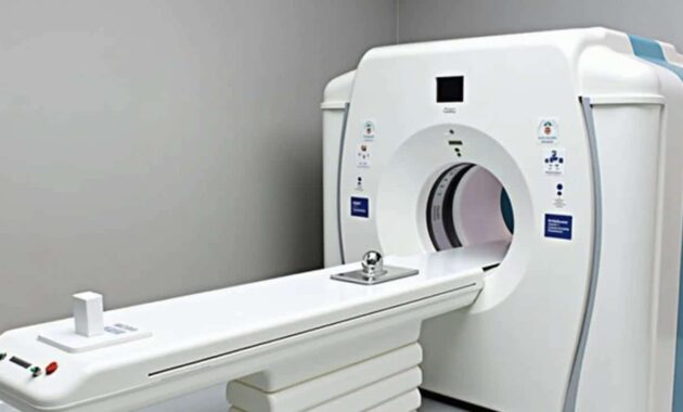
CT scans, or computed tomography scans, use X‑rays to produce detailed cross‑sectional images of the body. They are standard in emergency rooms, stroke units and cancer wards. Their use has grown so rapidly that, in the United States alone, an estimated 93 million CT scans were carried out in 2023, involving more than 62 million people.
That surge is not just a matter of convenience. CT scans help spot internal bleeding, small tumours and clots that plain X‑rays can miss. They guide biopsies and help surgeons plan operations. In many cases, they shift the odds of survival.
Researchers now estimate that today’s levels of CT use in the US could contribute to about 103,000 additional cancer cases over the lifetimes of those scanned.
If that projection holds and practices stay the same, CT‑related cancers could account for roughly 5% of all new annual cancer diagnoses in the country. That would place medical scanning in the same order of magnitude as some better-known lifestyle risks: alcohol consumption is linked to around 5.4% of cancers, and obesity to about 7.6%.
What science says about scanners and cancer
CT scanners emit ionising radiation. This energy can damage DNA and, in rare cases, trigger changes that lead to cancer years or decades later. The link is not hypothetical; it is based on long-term epidemiological data and risk models built from exposed populations, including survivors of Hiroshima and Nagasaki and patients who received diagnostic or therapeutic radiation.
The JAMA Internal Medicine analysis, based on more than 120,000 real clinical exams, did not track individual patients from scan to cancer diagnosis. Instead, it estimated risk using detailed information on the type of scan, area of the body imaged, number of phases (for instance, before and after contrast injection), patient age and sex. Those parameters were combined with established radiation risk models.
Even when the researchers applied more conservative assumptions, estimated cancer numbers linked to CT scans remained high.
There are uncertainties. Risk models extrapolate from higher doses to the lower doses used in modern imaging. People’s lifestyles, genetics and medical conditions also affect their personal cancer risk. Yet, across multiple scenarios, the trend was consistent: heavy reliance on CT imaging appears to leave a detectable mark on future cancer statistics.
➡️ Ritual of Seshen: the Egyptian-inspired lotus slips into your Mother’s Day routine
➡️ Pixie cut after 50: pro tricks to “knock 10 years off” when you go super short
➡️ Meet “stained glass hair”, the chic way to soften grey roots without hiding them
➡️ A mysterious radio signal heard in the universe is shaking up astrophysics
Who is most at risk from CT radiation?
Children and young patients
Radiation risk is not shared equally. Children are particularly vulnerable because their tissues are still developing and they have many years ahead in which a radiation‑induced cancer could emerge. The risk per scan is highest for babies, especially before the age of one.
For a single CT scan, a child may receive a similar radiation dose to an adult, yet their lifetime risk from that dose is significantly higher. That is why paediatric imaging protocols aim to “child‑size” the dose, and why many guidelines urge doctors to consider ultrasound or MRI first for non‑urgent problems in children.
High‑dose body regions
Risk also varies with the body part being scanned, because some regions require more radiation to produce a clear image. Abdomen, chest and pelvis exams sit near the top of the risk scale.
The JAMA‑based modelling suggests that abdominal and pelvic CT scans alone could account for more than 37,000 of the predicted cancers linked to current US scanning habits. Organs in these areas, such as the bowel, bladder and reproductive organs, are relatively sensitive to radiation.
- Abdomen: linked to potential cancers of the colon, liver and pancreas
- Pelvis: associated with bladder and reproductive cancers
- Chest: associated with lung cancer and, for women, breast cancer
- Neck and head: involve the thyroid and brain, with thyroid cancer and certain blood cancers a concern
Differences between men and women
The study found that certain cancers appear more frequently in women after radiation exposure, notably breast and thyroid cancers. Yet the average radiation dose women receive during CT scans is broadly similar to that given to men.
Biological differences and historical risk models suggest women carry a higher lifetime cancer risk from the same radiation dose than men.
Radiation risk estimates for women often come out higher because female breast and thyroid tissue are more sensitive to ionising radiation, and women tend to live longer, extending the window for a potential cancer to appear.
Balancing life-saving benefits and long-term risks
The numbers may sound alarming, but radiologists stress that CT scans prevent serious harm every day. The American College of Radiology, which sets key imaging standards in the US, points out that CT technology has helped cut hospital mortality by speeding up diagnoses and avoiding unnecessary surgery.
Stroke patients can receive clot-busting drugs faster because a CT scan can distinguish between a bleed and a blockage. Trauma teams use whole‑body CT to find hidden internal injuries within minutes. Cancer specialists rely on CT to stage disease accurately and plan targeted therapies.
No study has linked a specific CT scan in a named patient to a specific cancer; risk becomes visible only at the population level.
This does not mean the risk is imaginary. It means the extra cancers are spread thinly across millions of people. For one individual in an emergency, the choice is often clear: accept a small, delayed risk to gain a large, immediate benefit.
Rethinking imaging without losing diagnostic power
Public health agencies and medical societies are not calling for an end to CT scanning. The focus is on using scans more wisely and delivering the lowest dose that still produces a useful image.
Campaigns such as “Choosing Wisely” in the US and “Image Gently” for paediatric imaging encourage doctors to question routine or defensive scanning. They provide checklists and decision tools to help clinicians weigh radiation‑free alternatives, such as ultrasound or MRI, when these are clinically acceptable.
| Clinical situation | Common imaging choice | Lower‑radiation alternative |
|---|---|---|
| Suspected kidney stones in a young adult | CT abdomen/pelvis | Ultrasound first, CT only if needed |
| Chronic back pain without red flags | CT or MRI | No imaging initially; physiotherapy and review |
| Paediatric head injury | Head CT | Observation and clinical rules; CT only for higher‑risk cases |
Regulators are also tightening dose controls. Accreditation programmes for imaging centres require regular equipment checks, staff training and dose monitoring. Newer scanners can perform the same examination at far lower doses than machines from a decade ago, thanks to better detectors and image reconstruction algorithms.
How patients can question a proposed scan
Patients are not expected to calculate radiation risk on their own, but they can shape decisions by asking simple questions before agreeing to a CT scan, especially for non‑emergency problems.
“Do I really need this scan now, or could we wait or use a different test?” is often the single most powerful question.
Other useful questions include:
- “Is there an ultrasound or MRI option that would work instead?”
- “Have I had similar scans in the past few years, and do you need to repeat them?”
- “Does this imaging centre track and optimise radiation doses?”
For parents, it is reasonable to ask whether the scanner uses paediatric settings and whether the exam has been tailored to the child’s size. If the answer is vague, asking for a paediatric radiologist’s input can be justified.
Where artificial intelligence fits into safer imaging
AI is starting to influence both when scans are requested and how they are performed. Some hospitals are testing algorithms that flag potentially unnecessary repeat exams, such as multiple CT scans of the same region over a short period.
Other systems track the total radiation dose a patient receives across different clinics, giving doctors a clearer picture of cumulative exposure. This kind of “dose passport” could help clinicians pause before ordering yet another scan.
On the technical side, AI‑assisted reconstruction can clean up noisy images taken at lower radiation levels, allowing radiologists to interpret them confidently without turning up the X‑ray power.
Understanding radiation dose and risk in everyday terms
Radiation dose from imaging is usually expressed in millisieverts (mSv). A chest X‑ray might give about 0.1 mSv. A typical CT of the abdomen and pelvis might range from 5 to 10 mSv, sometimes more with multiphase studies. For comparison, the average person receives around 3 mSv a year from natural background radiation, mainly from the ground and cosmic rays.
An often-quoted way to think about risk is the “one in a thousand” rule of thumb. For a middle‑aged adult, a CT scan in the 10 mSv range might raise lifetime cancer risk by roughly 1 in 1,000. That is small for the individual, but scaled up across tens of millions of scans, the extra cancers add up.
Risk is also cumulative. Several CT scans over a few years, especially of high‑dose regions like the abdomen and chest, can push a person’s added risk higher. That does not mean previous scans were a mistake. It argues for keeping a record and making sure each new scan brings fresh value.
Practical scenarios where choices matter
Consider a 30‑year‑old with mild, recurring abdominal pain. In the absence of alarming signs such as fever, weight loss or blood tests pointing to a serious problem, some guidelines suggest starting with ultrasound and watchful waiting. Jumping straight to CT may generate beautiful images without changing treatment, while adding dose that could have been avoided.
Now think of a 70‑year‑old who arrives at A&E with chest pain and shortness of breath. Here, a CT pulmonary angiogram could promptly confirm or rule out a life‑threatening blood clot in the lungs. In that situation, the immediate benefit generally outweighs any small rise in long-term cancer risk.
The real challenge is not choosing between scans and no scans, but matching the right test to the right patient at the right moment.
As imaging technology advances and pressure grows to control healthcare costs, more attention is likely to fall on dose tracking, referral criteria and patient communication. That trend may not eliminate radiation risk from medical scanners, yet it can shrink it, while preserving—and sometimes enhancing—the life‑saving power of modern imaging.